
Atlases
Register mouse brain, rat brain and spinal cord sections, even when they are cut at an angle.
Mouse, rat and spinal cord atlases
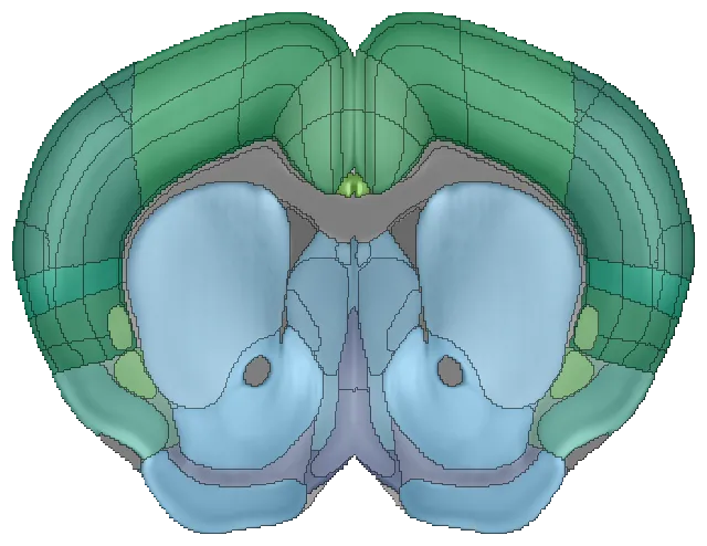
Allen Mouse Brain Atlas
The standard reference for mouse brain studies.
mouse brain · 643 regions · 25 µm
Wang et al. 2020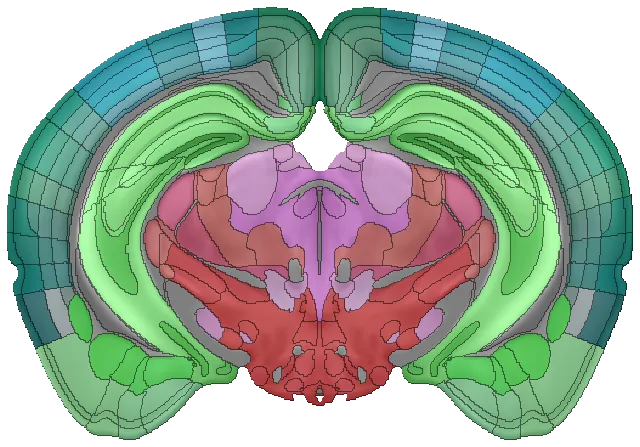
Kim Unified Mouse Brain Atlas
Finer subregions, with Allen and Franklin-Paxinos labels unified.
mouse brain · 1,001 regions · 20 µm
Chon et al. 2019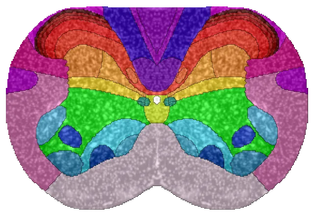
Allen Mouse Spinal Cord Atlas
Every segment from cervical to coccygeal.
mouse spinal cord · 73 regions · 10 µm
Fiederling et al. 2021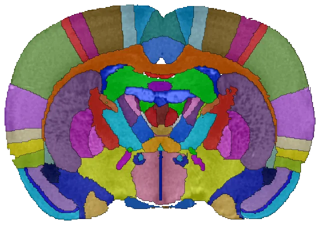
Waxholm Sprague Dawley Rat Brain Atlas
An open reference for the adult rat brain.
rat brain · 224 regions · 39 µm
Kleven et al. 2023
★ Our most popular feature
Make the atlas fit your tilted sections
Few cuts are perfectly coronal. At the surface of a mouse brain, a couple of degrees of tilt puts the two hemispheres at different AP positions, so a flat coronal atlas can't match both sides. NeuroQP lets you tilt the atlas to match the section.
- Adjust left/right and dorsal/ventral tilt, with a live preview in both viewers
- Set tilt once per animal; every slice keeps its own AP and landmarks
- Counts, areas, overlays and density plots all use the tilted geometry
Kim mouse atlas · target AP −1.30 mm from Bregma
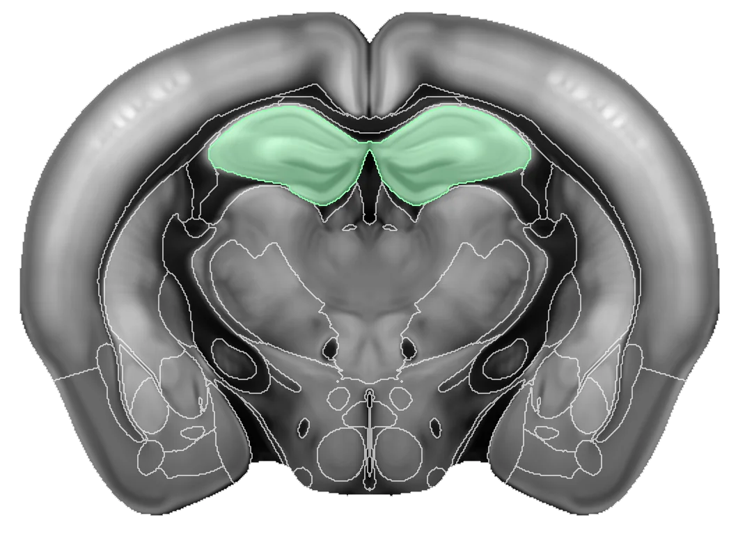
Left AP −1.30 · Right AP −1.30 mm
Left–right tilt 0°
Dorsal–ventral tilt 0°
Sketch angles exaggerated 2×
Spinal cord, segment by segment
Spinal cord sections get the same workflow as brain sections. Register each section to the Allen Mouse Spinal Cord Atlas, count cells by anatomical region, and compare across cervical, thoracic, lumbar and sacral levels.
- Name sections by segment (C1, T9, L3) instead of exact coordinates
- Dorsal horn, ventral horn and white matter as quantifiable regions
- Same detection, classifiers and statistics as brain projects
Allen mouse spinal cord atlas · 34 segments
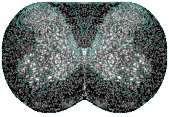

Left and right, separately or together
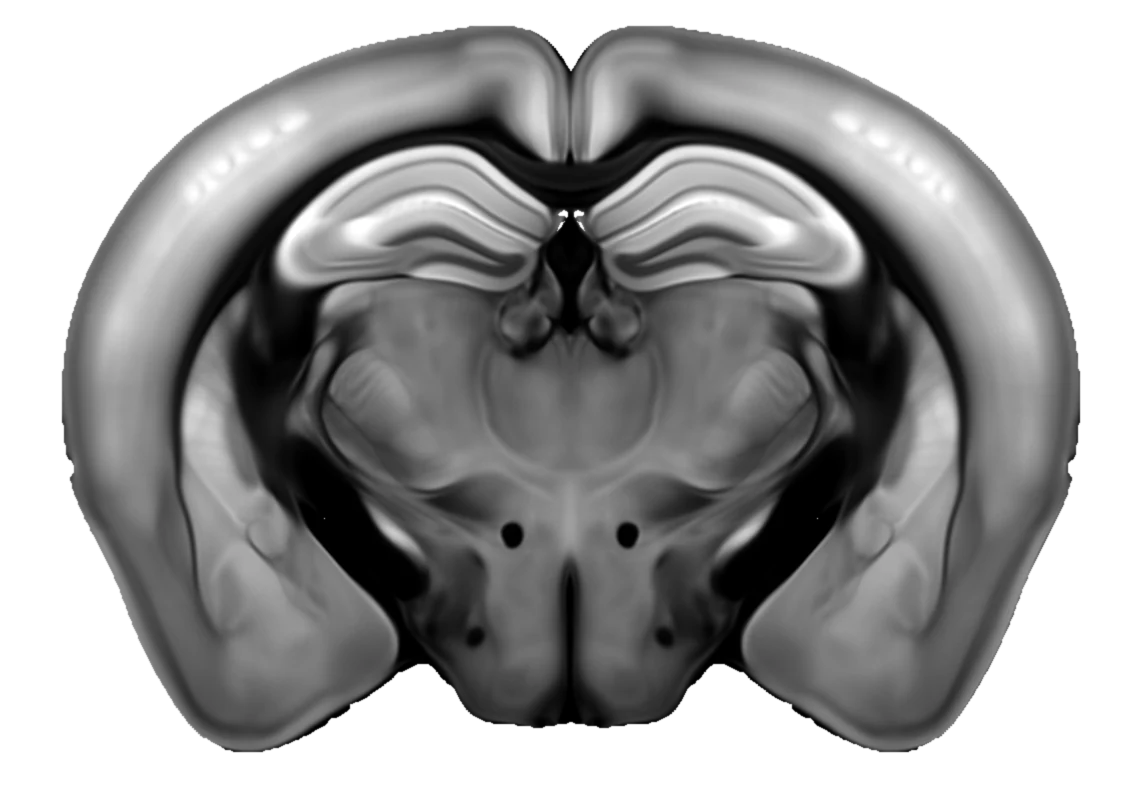
Lateralized injections, unilateral lesions and asymmetric effects are easy to quantify. Measure the regions where hemisphere matters separately and keep the rest joint.
- Separate left/right measurements in tables, viewers and plots
- Choose joint or separate for each region
Kim mouse atlas · AP −1.70 mm
Selected regions
Bring your tilted sections, spinal cords and rat brains.
Upload, review, and export region-level results without plugins, scripts, or a GPU.