
NeuroQP consulting: Let's talk.

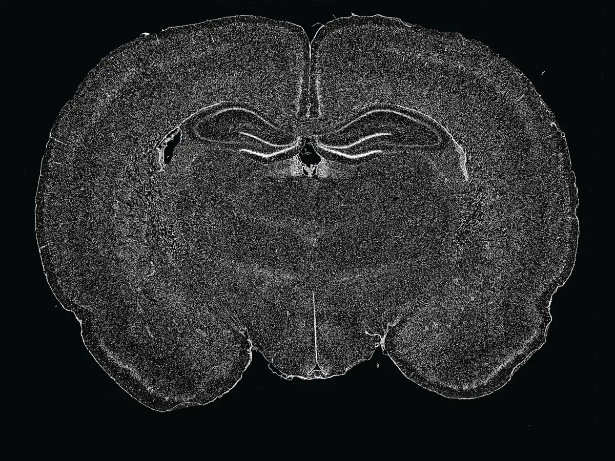
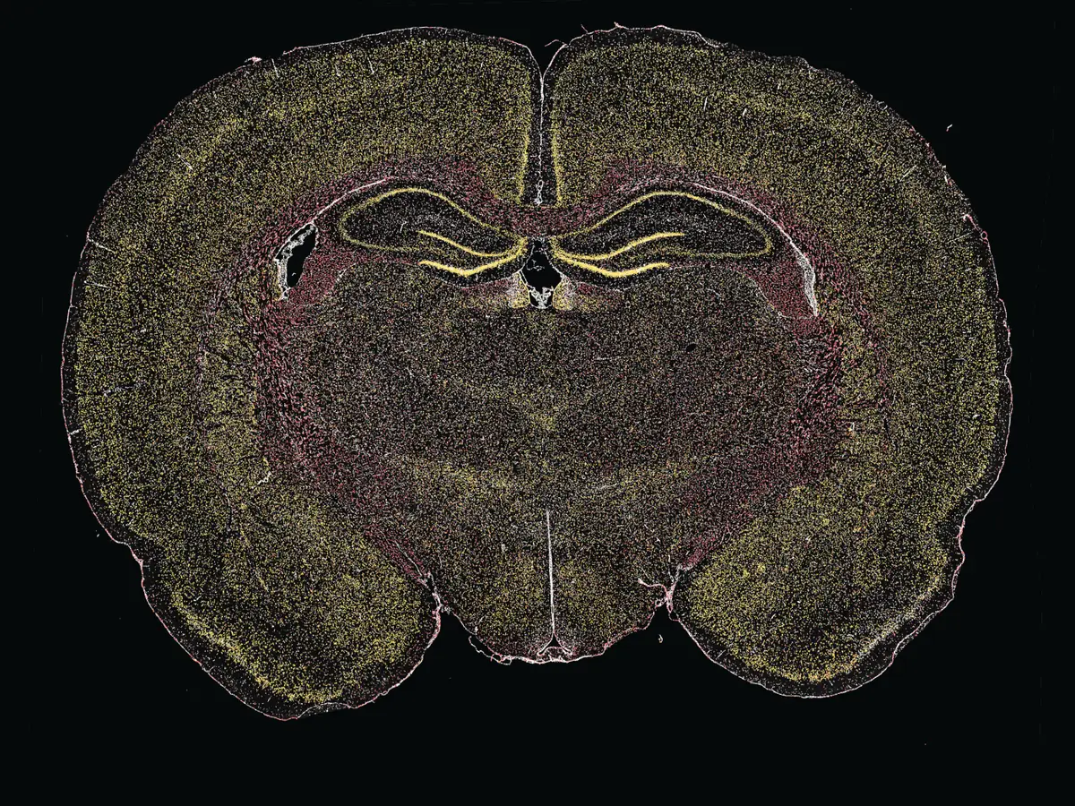
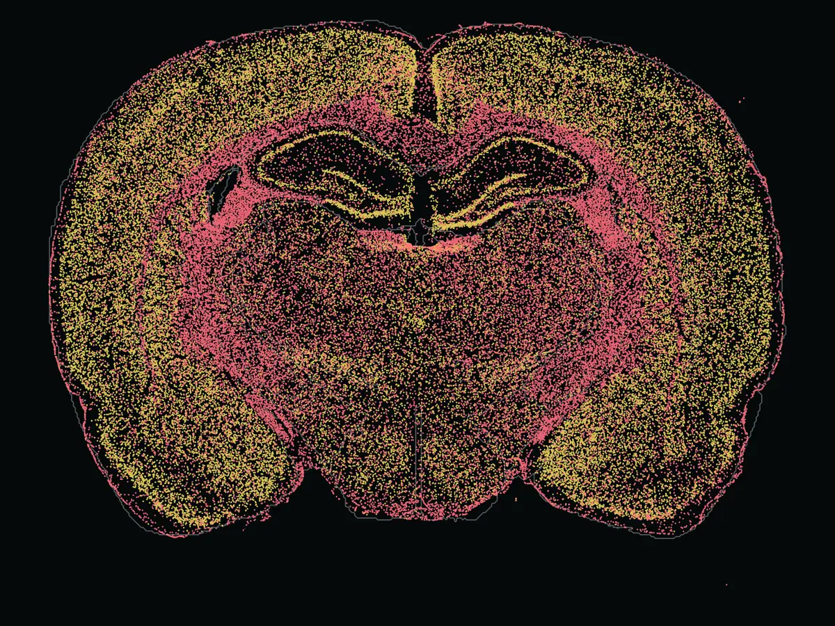
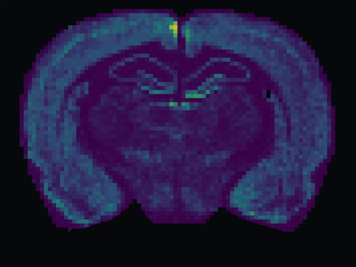
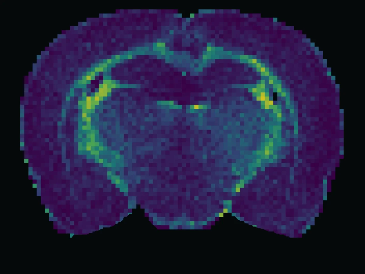
Coronal rat brain section stained with DAPI
DAPIRat coronal section, AP −3.12 mm
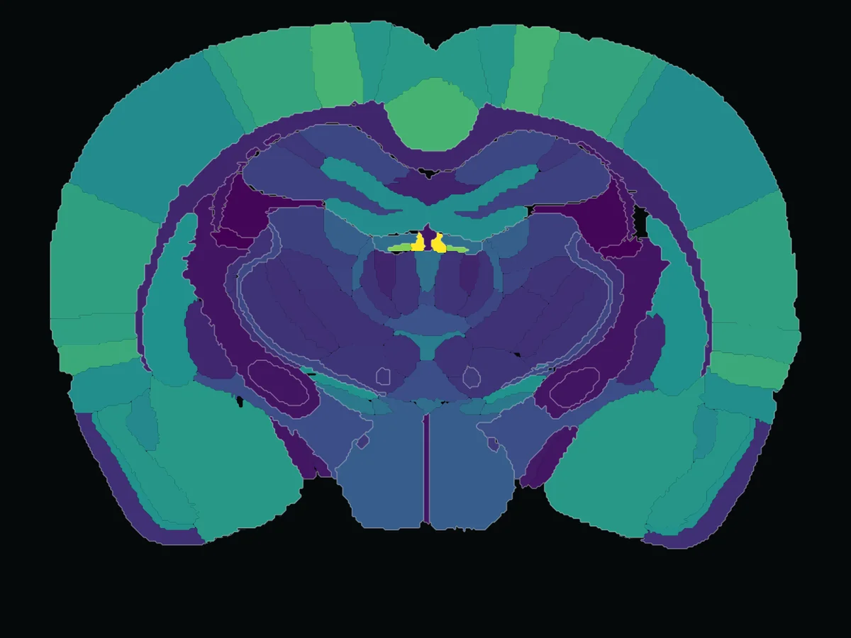
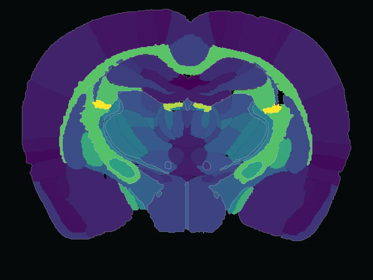
Rat microscopy images: Maric et al. (2021), CC BY 4.0, adapted with NeuroQP overlays.

Send us your brain or spinal cord images. We detect and classify the cells, map them to the atlas and hand back statistics and figures you can publish with confidence. Every analysis is tailored to your experiment and your question.

Species
Mouse, rat or yours
Tissue
Brain, spinal cord
Markers
DAPI, NeuN, Olig2, cFos, TRAP or yours

Two ways we can help

Complete project analysis. Done for you.

Send us your images. We hand back the finished analysis: statistics and figures ready for your paper. Follow the work live in NeuroQP and inspect every step we take.

  • Atlas registration
  • Cell counts
  • Quality control
  • Statistics
  • Figures
  • Live view in NeuroQP
Hand off my project

Custom analysis. Built for your question.

New stain, co-expression or an unusual question? We develop the algorithms and the analysis for your experiment, and hand back the statistics and figures.

  • Algorithm development
  • Cell classifiers
  • Co-expression
  • Statistics
  • Figures
Scope a custom analysis

From first email to figures

  1. Email us

    Your question, species, markers and image count.

  2. Talk it through

    A call about your project, so we understand exactly what you need.

  3. Get a proposal

    Scope, timeline and price, before any work starts.

  4. We analyze, you steer

    We run your analysis or build what it needs. You see results as they come in and steer the direction.

  5. Get your figures

    Plus the statistics and the data behind every number.

Talk to us about your project.

The email comes prefilled with the questions we need answered. We'll reply and set up a call.